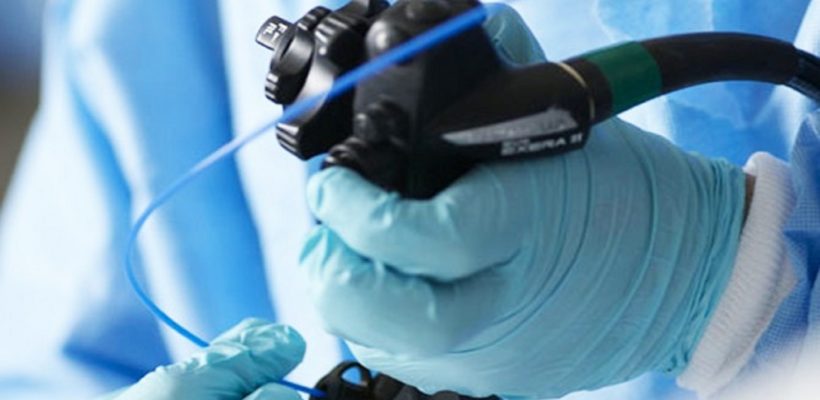
GI Endoscopy
Upper GI endoscopy
Upper GI endoscopy is a procedure that uses a flexible endoscope to visualize the upper GI tract. The upper GI tract includes the oesophagus, stomach, and duodenum – the first part of the small intestine.
Applications
-
Treating diffused mucosal bleed with Argon plasma coagulation (APC)
Dilation of narrowed food pipe (Balloon dilation of stricture and achalasia cardia)
Metallic stenting of narrowed segments of food pipe or stomach
Removal of polyps (polypectomy)
Creating alternative pathway for feeding directly to stomach (PEG) or small intestine (PEG-J)
Draining fluid collections though stomach (Cystogastrostomy) in patients with pancreatitis
How is upper GI endoscopy performed?
During the procedure, patients lie on their back or side on an examination table. An endoscope is carefully fed down the oesophagus and into the stomach and duodenum. A small camera mounted on the endoscope transmits a video image to a video monitor, allowing close examination of the intestinal lining. Air is pumped through the endoscope to inflate the stomach and duodenum, making them easier to see. Special tools that slide through the endoscope allow the doctor to perform biopsies, stop bleeding, and remove abnormal growths.
What problems can upper GI endoscopy detect?
Upper GI endoscopy can be used to determine the cause of
-
Abdominal pain
Nausea
Nomiting
Swallowing difficulties
Gastric reflux
Unexplained weight loss
Anaemia
Bleeding in the upper GI tract
It is used for both diagnostic and therapeutic procedures.
Diagnostic upper GI endoscopy is done to detect
-
Ulcers
Abnormal growths
Obstruction
Inflammation
Hiatal hernia
-
source of bleeding
tissue samples (biopsy) are also taken during endoscopy and sent for pathological examination to confirm the diagnosis
The following therapeutic (treatment) procedures are also performed through upper GI endoscopy:
-
Foreign body removal
Treat bleeding ulcers by
-
injection of medication (injection therapy)
application of heat (coagulation) or
application of clips (hemoclips) to the bleeding vessel
Treat bleeding varices (engorged veins in liver disease) by applying plastic rings (EVL)
Glue injection for gastric varix
Lower GI Endoscopy
Lower GI endoscopy allows your doctor to view your lower gastrointestinal (GI) tract. Your entire colon and rectum can be examined (colonoscopy). Or just the rectum and sigmoid colon can be examined (sigmoidoscopy).
During endoscopy, a long, flexible tube is used to view the inside of your lower GI tract.
Prep Instructions Before the exam
-
Follow these and any other instructions you are given before your endoscopy. If you don’t follow the doctor’s instructions carefully, the test may need to be cancelled or done over.
For a colonoscopy, you may be told not to eat and to drink only clear liquids for one to three days before the exam.
Take any laxatives that are prescribed for you. An enema may also be prescribed.
Arrange for someone to drive you home after the exam if you will be sedated.
Tell your health care provider before the exam if you are taking any medications, vitamins, supplements, or have any medical problems. Please bring a written medication list including or printed dosages.
The procedure
-
Colonoscopy can take 30 minutes or longer. Sigmoidoscopy often takes about 20 minutes.
You lie on the stretcher or bed on your left side.
For colonoscopy, you are given sedating (relaxing) medication through an IV line.
Sigmoidoscopy often doesn’t require sedation.
The endoscope is inserted into your rectum. You may feel pressure and cramping. If you feel pain, tell your doctor or nurse. You may receive more sedation which includes pain medication and an anti-anxiety medication or a mild anesthetic.
The endoscope carries images of your colon to a video screen. Prints of the images may be taken as a record of your exam.
When the procedure is done, you rest for at least 30 minutes. You may have some discomfort right after the procedure from trapped air. This can be relieved by changing position and passing the air. If you have been sedated, you must have an adult to drive you home.
